Figure 1.

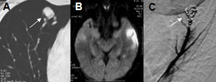
Radiological features of patient 1. A: Contrast-enhanced thoracic CT scan shows PAVMs (arrow); B: Brain MRI diffusion hyperintensity in the left temporal cortical region; C: Pulmonary angiography showing PAVMs after embolization (arrow).

Radiological features of patient 1. A: Contrast-enhanced thoracic CT scan shows PAVMs (arrow); B: Brain MRI diffusion hyperintensity in the left temporal cortical region; C: Pulmonary angiography showing PAVMs after embolization (arrow).