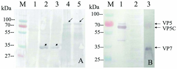
Figure 4.
Western blotting analysis of expressed proteins VP5, VP7 and GCRV. A: Western blotting analysis of expressed VP5 and VP7 cell lysate extracts in E. coli with native GCRV antibody. M, Standard protein marker; Lane 1, empty vector cell lysate extracts induced with IPTG for 3 h as negative control; Lanes 2-3, VP7 cell lysate extracts induced by IPTG for 3 h; Lanes 4-5, VP5 cell lysate extracts induced with IPTG for 3 h. The long arrowhead indicates VP5 and short arrowhead indicates VP7. B: Western blotting of purified GCRV with VP5Ab and VP7Ab; Lane 1, purified native GCRV particles with VP5Ab; Lane 3, purified native GCRV particles with VP7Ab; Lane 2, un-infected CIK cell as negative control.

