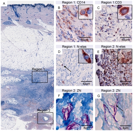
Figure 2. Histopathological presentation of secondary lesions.
Histological sections (nodule 2 of patient 2) were stained either with Ziehl-Neelsen (counterstain methylenblue; A, F, G) or with antibodies against cell surface or cytoplasmic markers (counterstain haematoxylin; B–E). A: Overview over excised tissue specimen revealing typical BU pathology features like fat cell ghosts, necrosis, epidermal hyperplasia and AFB (region 2) as well as a strong mixed infiltration typically observed in successfully treated BU lesions (region 1). B: CD14 staining of macrophages/monocytes; C: CD3 staining of T-cells; D: Elastase staining of neutrophils. In the necrotic region 2 large numbers of elastase-positive neutrophilic debris (E) and small clumps of AFB (F) with a beaded appearance (G) were observed.

