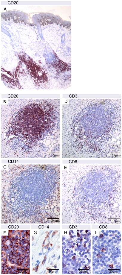
Figure 3. Presence of B-cell clusters in the secondary lesions.
A: Band of CD20 positive B-cells in sections of ulcer 2 of patient 1. B–E: serial sections of nodule 3 of patient 2 with a small dense cluster of CD20 positive B-cells (B) surrounded by CD14 positive macrophages/monocytes (C) and few interspersed CD3 positive T-cells (D) from which the majority was CD8 negative (E). Higher magnification (F–I) revealed a very dense package of the B-cells.

