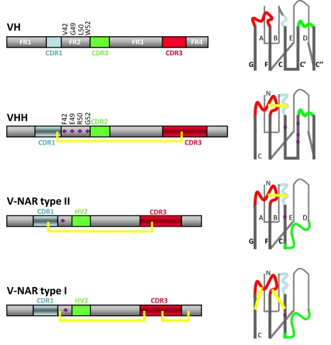
Figure 3. Schematic of the rearranged VH, VHH, and V-NAR type I or type II.
At left are displayed the linear sequence hallmarks such as the complementarity determining region (CDR) within the more conserved framework (FR) residues (gray). The disulfide bonds connecting the CDR3 with the CDR1 or FR in a VHH or V-NAR are shown by the yellow line. The folded structures of the V domains with the nine or seven β strands (named A to G in V-NARs, and A to G with insertion of strands C′ and C′′ between C and D for VH and VHH) forming two β-sheets are shown on the right. The purple diamonds on the structure denote the VHH hallmark residues in FR2 or the polar charged residues in the V-NARs. The N- and C-terminal ends of the polypeptides are shown in the structure of VHH and V-NAR type I.

