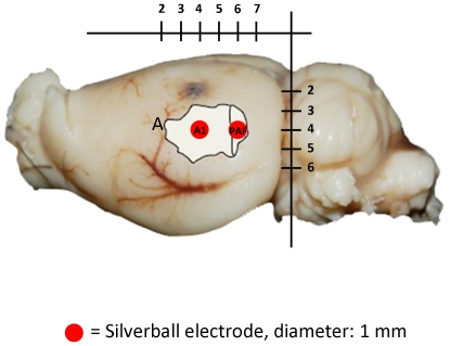
Figure 1. Electrode Placement.
Electrode placement (silverball electrodes) in primary auditory cortex (A1) and posterior auditory field (PAF) in auditory cortex (A). The anatomical labelling of auditory fields was taken from [38] and matched to a rat brain from our animals. The indicated scaling is in mm.

