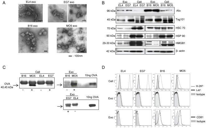
Figure 1. Characterization of tumor exosomes.
(A) EM micrographs of exosomes isolated from EL4, EG7, B16 and MO5 cell culture supernatants. (B) Western blot analysis of exosomes and cell lysates. 10 µg of proteins were loaded per lane. (C) IP detection of OVA protein (40∼45 kD) in both cell lysates and exosomes. (D) FACS analysis of MHC class I, MHC class II and CD81 expression on cells and exosomes.

