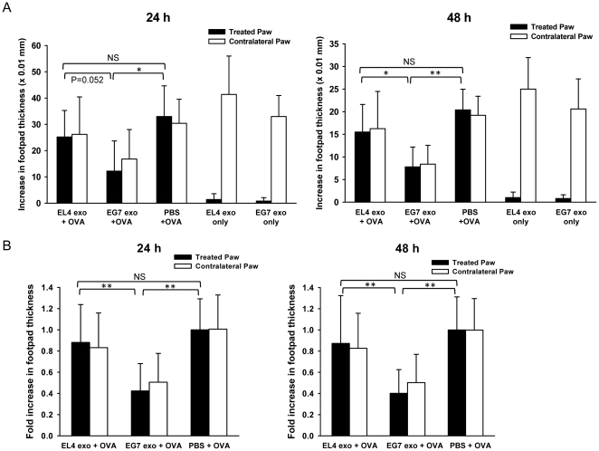
Figure 2. Suppression of OVA-specific DTH response by local administration of EG7 exosomes.
(A) Mice pre-sensitized with OVA were injected with 10 µg of EL4 exosomes plus 30 µg of OVA, 10 µg of EG7 exosomes plus 30 µg of OVA, 30 µg of OVA alone, 10 ug of EL4 exosomes alone or 10 ug of EG7 exosomes alone in 50 µl of PBS in their right hind paws. The left hind paws were all challenged with 30 µg of OVA in 50 µl of PBS. Paw swellings of both treated (right) and contralateral (left) paws were measured 24 h and 48 h post-challenge as the increase in footpad thickness (×0.01 mm). The results shown are from one representative experiment and are the means ± SD with n = 5. (B) The mean increase of footpad thickness of the treated paws in PBS group (OVA alone) at each time point was set to 1, and the increases of footpad thickness in EL4 exosomes plus OVA group and EG7 exosomes plus OVA group were normalized as fold increase. Figures show the pooled results of three independent experiments and are the means ± SD with n = 15. Significance at **: P<0.01; *: P<0.05; NS: not significant.

