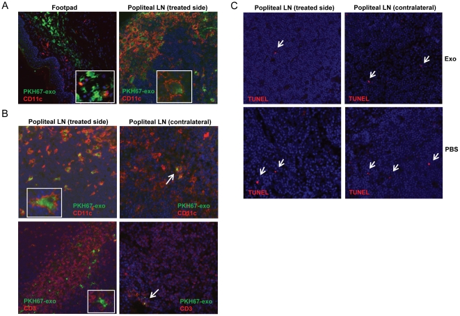
Figure 5. Exosome in vivo trafficking in DTH model.
PKH67-labeled exosomes were injected in the right footpad of OVA-sensitized mice as in the DTH experiment. Footpads and the popliteal LNs were harvested, cryo-sectioned and examined by immunofluorescence. Similar observations were made with different tumor exosomes and data show the representative figures of MO5 exosomes. (A) 24 h post-injection, exosomes (green) were captured by dermal CD11c+ cells (red) in footpads and transported to the treated-side LN. (B) 48 h post-injection, large numbers of exosome-internalized CD11c+ cells (red, upper left panel) appear in the treated-side LN. Exosomes (or exosome-containing cells) were also physically adjacent to CD3+ T cells (red, lower left panel). Only very few exosomes were observed in the contralateral LN. (C) TUNEL staining for apoptotic cells (red) in both side LNs 48 h post-injection. Magnification: 20×.

