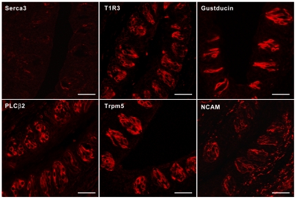
Figure 4. Normal taste bud morphology and expression of taste-signaling molecules in Serca3 KO mice.
Immunofluorescence images of Serca3-KO mouse circumvallate taste tissue sections with antibodies against Serca3, T1R3, α-gustducin, PLCβ2, Trpm5, and neural cell adhesion molecule (NCAM) indicate that Serca3 protein was absent in taste buds, whereas the expression of other proteins appeared unaltered. Scale bars, 25 µm.

