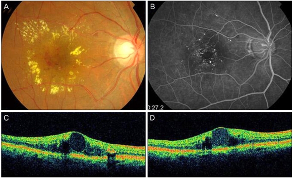
Fig. 1.
Group 1A idiopathic juxtafoveal retinal telangiectasis (case 12). (A) Color fundus photography of the left eye on presentation showed a lipid exudate with aneurysm-like capillaries in its center. (B) Fluorescein angiography of the left eye on presentation showed telangiectasis and microaneurysm. (C) Optical coherence tomography of the left eye on presentation indicates macular edema with foveolar detachment. (D) Macular edema showed no improvement 3 months after a repeat intravitreal bevacizumab treatment and focal laser photocoagulation.

