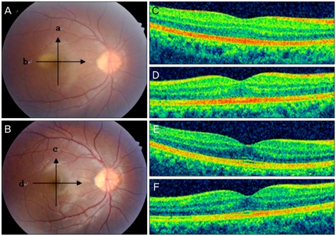
Fig. 1.
Case 2. (A) Fundus photograph showing retinal opacity on the day of trauma. (B) Fundus photograph at 7 days post-trauma showing resolution of commotio retinae. (C) and (D) are optical coherence tomography (OCT) images taken on the day of trauma, showing increased reflectivity in the area of the photoreceptor outer segment (these scans correspond to line scans a and b on A). (E) and (F) are OCT images taken 7 days post-trauma, revealing restoration to the normal alternated layer (these scans correspond to line scans c and d on B). Arrows denote the orientation of the OCT line scan.

