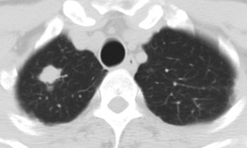
Figure 2b:
Necrotizing granuloma found in a 51-year-old man at pre-employment screening. (a) Posteroanterior chest radiograph shows an ill-defined right apical mass (arrow) obscured by overlying body structures. (b) CT image shows the irregular mass in the right upper lobe. The diagnosis was made on the basis of tissue obtained with CT-guided aspiration biopsy. Pathologic analysis revealed the mass was consistent with necrotizing granulomas (negative for acid-fast bacilli), and Mycobacterium kansasii grew on culture.

