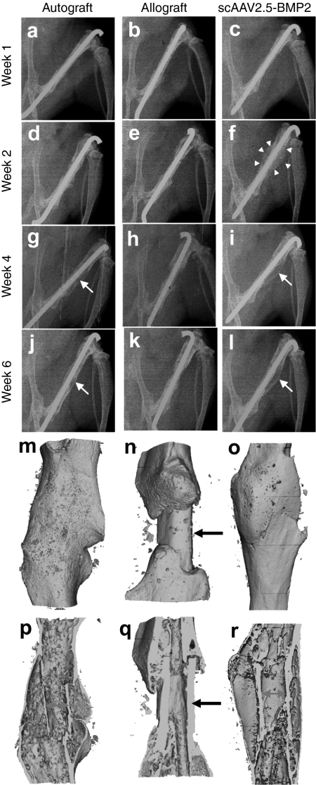
Figure 3.
Radiographic healing of murine femoral autografts, allografts, and self-complementary AAV serotype 2.5 vector (scAAV2.5)-bone morphogenetic protein-2 (BMP2)-coated allografts. (a–l) Longitudinal plane X-rays were obtained of the grafted femurs, and representative radiographs from an individual mouse taken at the indicated time after surgery from each group (n = 5) are shown. Note the remarkable amount of callus formed around the scAAV2.5-BMP2-coated allograft by week 2 (arrowheads in f), which remodels to form a new bone collar similar to that of autograft at week 4–6 (arrows in g,i,j,l). Representative 3D reconstructed micro-computed tomography (CT) images of the grafted femurs at 6 weeks are shown with (m–o) surface and (p–r) medial slice views to illustrate the indistinguishable new bone collar that completely surrounds (m,p) autografts and (o,r) scAAV2.5-BMP2-coated allografts, versus the lack of new bone formation around the medial segment of unremodeled allografts (arrows in n,q). Also of note is the extensive remodeling of autograft at this time, which is evident from the very thin cortical bone that is discontiguous with the host cortical bone, versus the largely unremodeled scAAV2.5-BMP2-coated allograft that is osteointegrated at both proximal and distal graft-host junctions p versus r).

