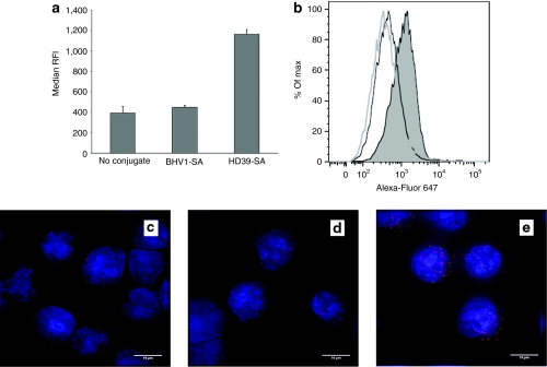
Figure 6.
Internalization of polymeric micelles containing Alexa-Fluor 647-labeled small interfering RNA (siRNA) with and without monoclonal antibody-streptavidin conjugate (mAb-SA) conjugate. DoHH2 lymphoma cells were incubated at 37 °C for 30 minutes with polymeric micelles bearing HD39-SA, BHV1-SA, or no conjugate then washed extensively with phosphate-buffered saline (PBS) then acid wash to remove surface based polymeric micelles prior to measuring fluorescence intensity by flow cytometric analysis. (a) The median relative fluorescence intensity (RFI) + s.d. of triplicate samples is shown. (b) A representative histogram depicting the relative fluorescence of DoHH2 cells treated with polymeric micelles bearing HD39-SA (shaded peak), BHV1-SA (black line, unshaded peak) or no conjugate (gray line, unshaded peak) is shown. Treated cells were cytospun and stained with DAPI nuclear stain (blue fluorescence). Representative random images captured from cells treated with polymeric micelles bearing no conjugate (c), nontargeting BHV1-SA (d), or CD22-targeted HD39-SA (e) are shown. Bar = 10 µm.

