Figure 4.
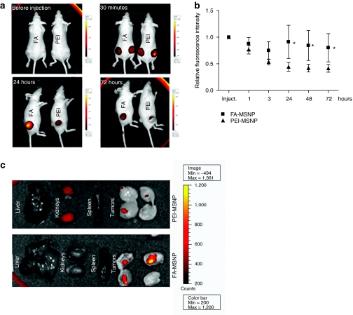
Tumor accumulation and retention of mesoporous silica nanoparticles (MSNPs) through folate (FA)-functionalization. (a) FA-conjugated and nonconjugated particles (PEI) were injected peritumorally and followed by the IVIS Lumina II imaging system for 72 hours. Scale bars range from 300 to 8000 counts for images 30 minutes to 72 hours, whereas scale bar is 200 to 4500 counts for control image (before injection). (b) Graph shows the quantification of the fluorescence intensity in tumors injected with FA-MSNPs and PEI-MSNPs over time as related to the initial intensity within each tumor (n = 6, two tumors per animal). (*P < 0.05). (c) Ex vivo imaging of internal organs and tumors 72 hours after injection of MSNPs. The tumors were cut in half to visualize particles within the tumor tissue.

