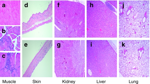
Figure 6.
Histopathological examination of the treated animals. Animals were treated as described in Figure 5. Organs were dissected 24 hours after treatment and fixed in formalin, embedded in paraffin, and stained with eosin and hematoxylin. Images were taken on the Olympus BX45 microscope with a Sony DXC-S500 digital camera. Histopathological sections of: (a) muscle tissue of the negative control, (b) plasmid-treated and (c) stearyl-transportan 10 (TP10)/plasmid treated animals; (d) skin after treatment with plasmid or (e) stearyl-TP10/plasmid complexes; (f) kidney after treatment with plasmid or (g) stearyl-TP10/plasmid complexes; (h) liver after treatment with plasmid or (i) stearyl-TP10/plasmid complexes; (j) and lung after treatment with plasmid or (k) stearyl-TP10/plasmid complexes.

