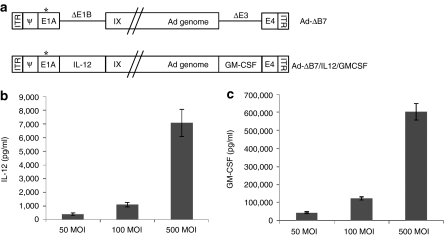
Figure 1.
Characterization of oncolytic adenovirus coexpressing interleukin (IL)-12 and granulocyte-macrophage colony-stimulating factor (GM-CSF). (a) Schematic representation of the genomic structures of adenovirus (Ad) Ad-ΔB7 and Ad-ΔB7/IL12/GMCSF. Ad-ΔB7 contains mutated E1A (open star, mutation at Rb protein–binding site), but lacks E1B 19 and 55 kDa (ΔE1B), and E3 region (ΔE3); the murine IL-12 and murine GM-CSF were inserted into E1 and E3 region of Ad genome, respectively. The level of IL-12 (b) and GM-CSF (c) expression was confirmed in B16-F10 cells after infection with Ad-ΔB7/IL12/GMCSF at different MOIs. Cell culture supernatants were collected at 48 hour after infection, and the level of IL-12 and GM-CSF was quantified by conventional enzyme-linked immunosorbent assay kit. Data represent the mean ± SE of triplicate experiments, and similar results were obtained from at least three separate experiments. MOI, multiplicity of infection.

