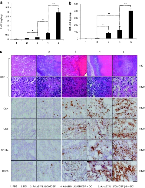
Figure 3.
Generation of antitumor immune response by Ad-ΔB7/IL12/GMCSF in combination with dendritic cell (DCs). The level of interleukin (IL)-12 (a) and granulocyte-macrophage colony-stimulating factor (GM-CSF) (b) expression in tumor tissues treated with phosphate-buffered saline (PBS), DCs, Ad-ΔB7/IL12/GMCSF, Ad-ΔB7/IL12/GMCSF plus DCs, or Ad-ΔB7/IL12/GMCSF (H; high-dose of Ad) plus DCs. (c) Histological and immunohistochemical analysis in tumor tissues treated with Ad-ΔB7/IL12/GMCSF and/or DCs. Tumor tissues were collected from mice at 3 days after final treatment, and paraffin section of tumor tissue was stained with hematoxylin and eosin (H&E) (top two rows, original magnification: ×40 and ×400). Immune cells infiltrated in tumor tissues were examined by anti-CD4 antibody (third row), anti-CD8 antibody (fourth row), anti-CD11c antibody (fifth row) and anti-CD86 antibody (bottom row). Original magnification: ×400. Data points represent the mean ± SE of at least three mice per group, and similar results were obtained from at least three separate experiments. *P < 0.05; **P < 0.01.

