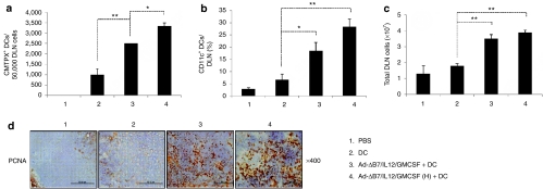
Figure 5.
Dendritic cells (DC) migration to the draining lymph nodes (DLNs). Ex vivo generated DCs were labeled with CMTPX. Two days after final treatment, single cells were collected from DLNs and the migration was quantified by fluorescence-activated cell sorting analysis. (a) The number of CMTPX+ DCs in DLNs from mice treated with phosphate-buffered saline (PBS), DC, Ad-ΔB7/IL12/GMCSF plus DCs, or Ad-ΔB7/IL12/GMCSF (H; high-dose of Ad) plus DCs were quantified on a fluorescence-activated cell sorter, and data from 50,000 events were represented. (b) CD11c+ DCs in the DLNs from mice treated with Ad-ΔB7/IL12/GMCSF and/or DCs. (c) Total cell number of DLNs from different groups of mice. Data points represent the mean ± SE of triplicate experiments. All results in this figure represent results of at least three mice per group, and similar results were obtained from at least two separate experiments. *P < 0.05; **P < 0.01. (d) Immunohistochemical staining of proliferating cell nuclear antigen in draining lymph node tissues. Original magnification: ×400. GM-CSF, granulocyte-macrophage colony-stimulating factor; IL, interleukin.

