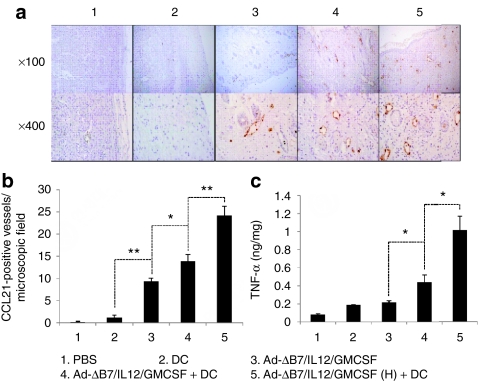
Figure 6.
Visualization and quantification of CCL21+ lymphatic vessels in tumors treated with Ad-ΔB7/IL12/GMCSF and/or dendritic cells (DCs). Tumors harvested from mice treated with phosphate-buffered saline (PBS), DC, Ad-ΔB7/IL12/GMCSF, Ad-ΔB7/IL12/GMCSF plus DCs, or Ad-ΔB7/IL12/GMCSF (H; high-dose of Ad) plus DCs were embedded in paraffin and sectioned. (a) Immunohistochemical staining of CCL21+ lymphatic vessels in tumor tissues. Original magnification: ×100 & ×400. (b) The means of CCL21+ lymphatic vessel in microscopic field (×200). (c) Tumor necrosis factor (TNF)-α expression in the tumor tissues was measured by conventional enzyme-linked immunosorbent assay. Data points represent the mean ± SE of triplicate experiments. All results in this figure represent results of at least three mice per group, and similar results were obtained from at least two separate experiments. *P < 0.05; **P < 0.01. GM-CSF, granulocyte-macrophage colony-stimulating factor; IL, interleukin.

