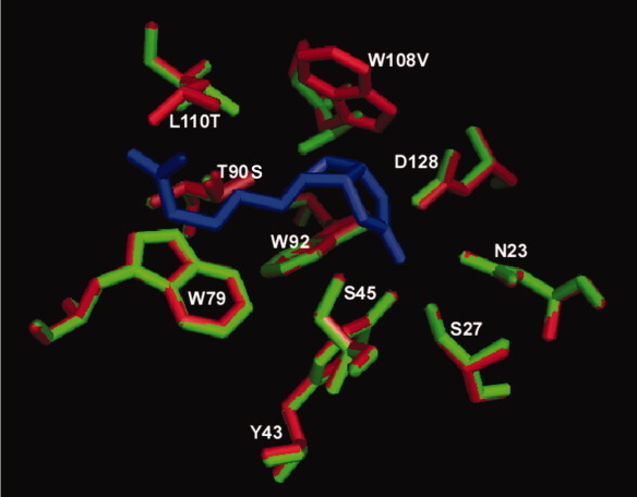
Figure 2.

Comparison of binding pocket residues between wild-type (red) streptavidin and R7-2 (green). Biotin is represented as dark blue sticks. The position of most residues remained virtually identical to the wild-type protein, including the backbone residues of the active site mutations W108V and L110T that produced a broadening effect on the binding pocket of the selected variants.
