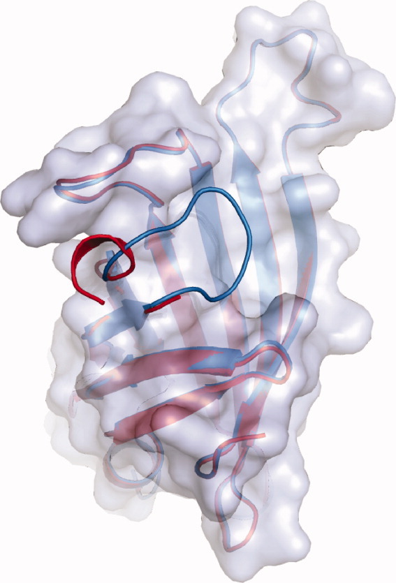
Figure 4.

Comparison of the overlaid loop structures of variants R4-6 and R7-2 (both in gray). The structures were derived from protein crystals formed in the absence of any ligand (structures R4-6 [2] and R7-2 [6]). The R4-6 loop (red) is open disordered and only partially visible. The R7-2 loop (blue) is closed and ordered.
