
Figure 3.
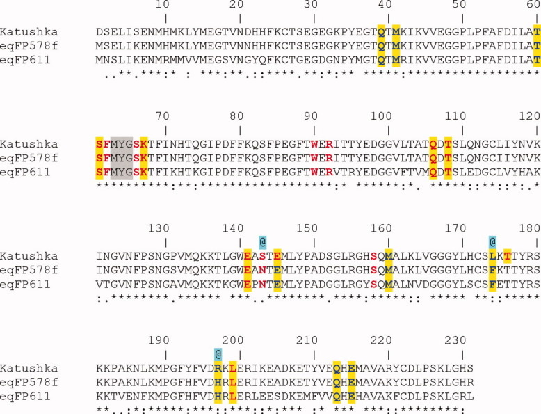
Sequence alignment displaying the residues interacting with the cis/trans chromophores. Red – direct H-bonding, Red (highlighted in yellow) – water mediated H-bonding, Blue – (highlighted in yellow) van der Waals contacts, @ – residues apparently responsible for the far-red shift in Katushka relative to eqFP578f and eqFP611, MYG – chromophore forming triad.