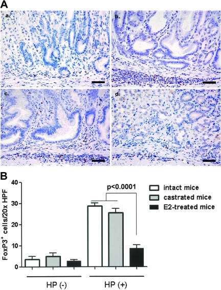
Fig. 4.
Quantification of FoxP3+ TREG cells in the gastric corpus of INS-GAS male mice at 16 weeks postinoculation (a–d). Representative high magnification images demonstrating immunohistochemical staining for FoxP3, a regulatory T-cell marker, in the gastric corpus in uninfected mice (a), H.pylori-infected intact mice (b) H.pylori-infected castrated mice (c) and H.pylori-infected and E2-treated mice (d). (e) Mean number of FoxP3+ cells in the gastric mucosa and submucosa assessed from 10 well-oriented fields (magnification, ×200; 1.00 mm2) per mouse gastric corpus. Bar, 40 μm.

