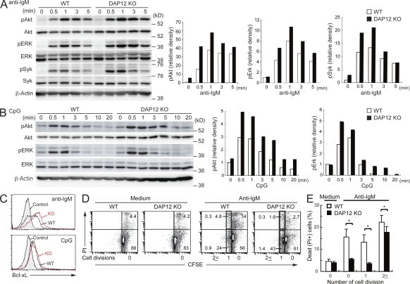
Figure 3.
Enhanced tyrosine phosphorylation of Erk, Akt, and Bcl-xL and prolonged survival in DAP12-deficient B cells. Purified B cells from the spleens of WT and DAP12-deficient mice were stimulated with 5 µg/ml of F(ab’)2 fragments of anti–mouse IgM (A and C) or 0.06 µM CpG (B and C). (A and B) The stimulated cells were lysed and then analyzed by immunoblotting with antibodies specific for indicated proteins. Bar graphs show the relative amount of each phosphorylated protein, as determined by densitometry, before and after stimulation. (C) The purified B cells 24 h after stimulation were fixed and stained with anti–Bcl-xL and then analyzed by flow cytometry. (D and E) The purified B cells were labeled with CFSE, stimulated with 10 µg/ml anti–mouse IgM or unstimulated (medium) for 48 h, stained with PI and analyzed by flow cytometry (D). The dead cell population was calculated as follows: PI+ cell frequency/PI+ and PI− cell frequency in each division peak of CFSE dilution (E). *, P < 0.05. n = 4. Data are representative of five (A), three (B), two (C), and four (D) independent experiments. Error bars show SD.

