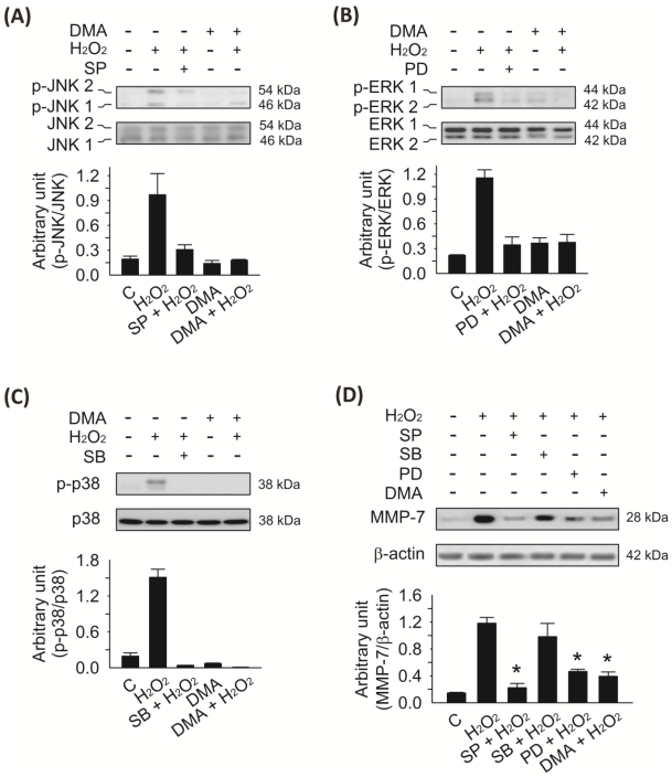
Figure 4.
Analysis of the different MAPK pathways activated by H2O2 in SW620 cells. SW620 cells were cultured in serum-free L-15 medium for 24 h to synchronize the cells and to make them quiescent prior to treatment with SP600125 (50 μM; JNK1/2 phosphorylation inhibitor), PD98059 (50 μM; ERK1/2 phosphorylation inhibitor), or SB203580 (10 μM; p38 MAPK phosphorylation inhibitor) for 30 min prior to the addition of H2O2 (5 μM) for 1 h. Cell lysates were prepared and immunoblotted with the antibodies indicated in the panel. A, the levels of phospho-JNK1/2 were evaluated by H2O2 stimulation and suppressed by SP600125 or DMA (100 μM) pretreatment. B, the levels of phospho-ERK1/2 were evaluated by H2O2 stimulation and suppressed by PD98059 or DMA (100 μM) pretreatment. C, the level of phospho-p38 MAPK was evaluated by H2O2 stimulation and suppressed by SB203580 or DMA (100 μM) pretreatment. Ratios of bands with phosphospecific versus non-phosphospecific MAPK antibodies were determined, and quantitated by densitometry using the ImageJ program (NIH). Means ± SD are given. D, H2O2-enhanced MMP-7 protein expression was associated with increased JNK and ERK signaling in SW620 cells. Pretreatment with DMA (100 μM), SP600125 (50 μM), or PD98059 (50 μM) for 30 min prior to H2O2 (5 μM) induction for 8 h in SW620 cells significantly declined the expression of MMP-7 compared with H2O2-only treatment (*p < 0.05). However, the level of MMP-7 protein was only slightly decreased with no significant difference effect in the presence of SB203580 (10 μM) after H2O2-induced MAPK stimulation. The bars in the lower panel denote means ± S.D. of three experiments for each condition determined from densitometry relative to β-actin.

