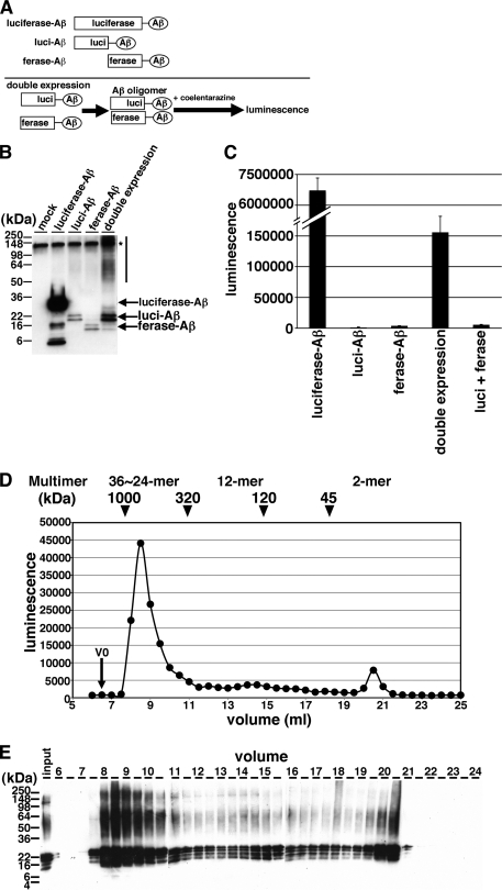
FIGURE 1.
Detection of Aβ dimer/oligomers using split-luciferase complementation assay. A, scheme of split-luciferase complementation assay for detection of Aβ oligomers. We generated split Gluc-tagged Aβ. Once Aβ forms oligomer, split Gluc proteins are reconstituted and show luminescence. Transient transfection of luciferase-Aβ, luci-Aβ, ferase-Aβ, or double (luci-Aβ and ferase-Aβ) cDNA into HEK293 cells. B, immunoblotting of CM from transfected HEK293 cells by anti-human Aβ monoclonal antibody 6E10. Asterisk shows nonspecific band. C, luminescence from the CM from transfected HEK293 cells. The luminescence ± S.D. in three independent experiments are shown. D, separation of Gluc-tagged Aβ in the CM from stably double (luci-Aβ/ferase-Aβ)-expressing HEK293 cells by size exclusion chromatography. V0 shows void volume. Calculated molecular masses are shown above the panel (arrowheads). E, samples eluted from 6–24 ml were analyzed by immunoblotting with anti-Aβ mAb 6E10.

