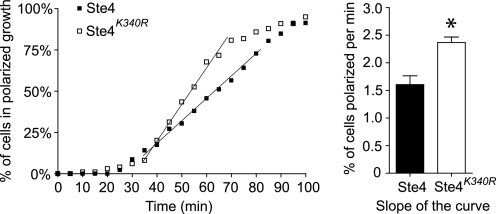
FIGURE 8.
Ubiquitination of Ste4 limits pheromone-induced polarized growth. Cells expressing either wild-type Ste4 or Ste4K340R were grown to A600 0.2 and treated with dimethyl sulfoxide (1%, final concentration) for 1.5 h before the addition of nocodazole (15 μg/ml in 1% dimethyl sulfoxide, final concentrations) for 2.5–3 h. Cultures were visualized by microscopy to confirm G2/M arrest. The cells were then loaded into a microfluidics device, and the nocodazole was washed out for 20 min prior to stimulation with 150 nm α factor. Cells were imaged at 5 min intervals for 2 h. For each cell, the time at which the first sign of polarized growth detectable in the differential interference contrast image was recorded and plotted as a function of time. The slope was taken from the linear portion of each curve, and the average slopes from three independent experiments are shown in the bar graph (right panel). The difference between Ste4 and Ste4K340R was statistically analyzed by t test. *, p < 0.05). Error bars, mean ± S.E.

