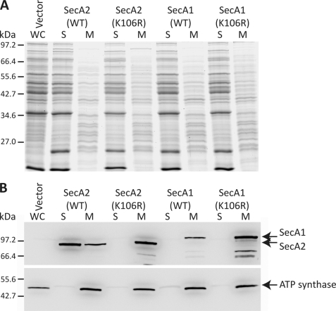
FIGURE 6.
Subcellular localization of SecA1 and SecA2. Mid-log cultures of C. difficile strain 630 harboring pMTL960 (Vector), pRPF186 (WT SecA2), pRPF187 (SecA2(K106R)), pRPF193 (WT SecA1), or pRPF194 (SecA1(K106R)) were induced with ATc (500 ng/ml) for 90 min. A, Coomassie Blue-stained SDS-polyacrylamide gel showing whole cell lysate (WC), soluble (S), and membrane (M) protein fractions. B, the location of SecA1 and SecA2 was detected using anti-Strep-tag II antibody. The integral membrane protein ATP synthase was used as a control for cell fractionation.

