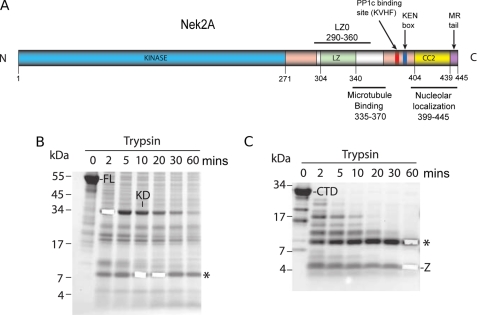
FIGURE 1.
Nek2 domain organization and limited proteolysis. A, scheme of full-length Nek2A kinase annotated with functional and structural motifs indicated with their position in the sequence. B, SDS-PAGE and Coomassie Blue analysis of full-length Nek2A kinase subjected to limited proteolysis for the times indicated (mins) FL, full-length protein; KD, kinase domain; *, 8 kDa fragment. C, as for B, but using the C-terminal non-catalytic region in the proteolysis assay. CTD, C-terminal domain; *, 8 kDa fragment; Z, nonspecific fragment corresponding to a region of Nek2A lacking tryptic sites.

