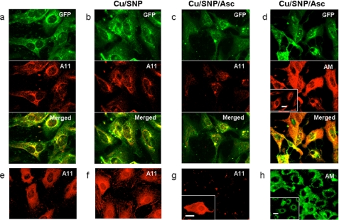
FIGURE 7.
Effect of ectopic expression of GFP-Gpc-1 and/or copper-NO supplementation on Aβ A11 immunoreactivity in untreated or ascorbate-treated Tg2576 fibroblasts. The immunofluorescence microscopy images show GFP-Gpc-1-transfected (a–d; green) or untransfected Tg2576 fibroblasts (e–h) that were untreated (a and e), exposed to medium containing 0.1 mm CuCl2 and 1 mm sodium nitroprusside (Cu/SNP) for 1 h (b and f), or exposed to medium containing 0.1 mm CuCl2 and 1 mm sodium nitroprusside for 1 h and then with fresh medium containing 1 mm ascorbate (Cu/SNP/Asc) for 3 h (c, d, g, and h) followed by staining for Aβ oligomers using the polyclonal antibody A11 (red) (a–c and e–g) or anMan-containing products using mAb AM (d (red) and h (green)). The inset in g shows A11 staining of untransfected cells that were incubated in fresh medium for 3 h after sequential exposure to CuCl2, sodium nitroprusside, and ascorbate. The insets in d and h show staining of untreated GFP-Gpc-1-transfected (d) or untransfected cells (h) for anMan-containing products using mAb AM. Scale bars, 20 μm.

