FIGURE 6.
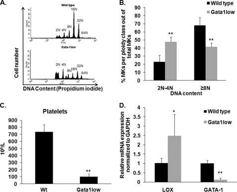
LOX levels in MKs of GATA-1low mice. A, ploidy analysis of wild-type and GATA-1low MKs. Cells were stained with CD41-FITC antibody and propidium iodide (DNA). Flow cytometry was performed on a BD Biosciences Calibur using CellQuest. Representative histograms are shown per group (wild-type, GATA-1low male littermates). B, quantification of MK percentage (CD41-positive cells) of diploid-tetraploid and polyploid fractions. C, platelet numbers of wild-type and GATA-1low MKs. Blood was collected via heart puncture and platelet number was assessed on a Hemavet blood analyzer. D, qRT-PCR on isolated MKs to evaluate LOX and GATA-1 gene expression. Data were normalized to GAPDH mRNA. Standard deviation bars represent the mean of five independent experiments; *, p < 0.05; **, p < 0.001.

