FIGURE 8.
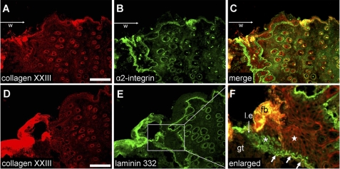
Distribution of collagen XXIII in skin wounds: collagen XXIII red (A and D), integrin α2β1 green (B), and laminin 332 green (E). Immunohistochemisty was performed on cryosections of wounds of 6-week-old mice (collected 4 days after wounding). The wound areas (w) are located at the left sides of the images, and the keratinocytes in the leading edges (l.e.) are moving from the right to the left. Co-staining was accomplished using sequential incubation with a polyclonal guinea pig anti-collagen XXIII antibody (red) followed by incubation with either a monoclonal antibody against the α2 integrin subunit (green) or a polyclonal rabbit anti-laminin 332 antibody (green). B and C, strong α2 integrin subunit staining can be detected at the wound edge (left sides of the pictures). As previously shown, the staining is most prominent at the wound edge. In the nonwounded areas and in the keratinocytes of the hair follicles, the signals are much weaker. D and F, collagen XXIII staining can be detected in all keratinocyte cell layers at cell-cell junctions. Near the wound edge, collagen XXIII was not present at cell-cell junctions (white asterisk); however, intracellular staining for laminin 332 can be seen. In the area (E, F), where a newly formed basement membrane was detected by immunofluorescence staining (arrows), intracellular staining for laminin 332 disappears, whereas cell-cell staining for collagen XXIII becomes apparent (white star). l.e., leading edge; w, wound area; gt, granulation tissue; fb, fibrin clot. Bar, 100 μm.

