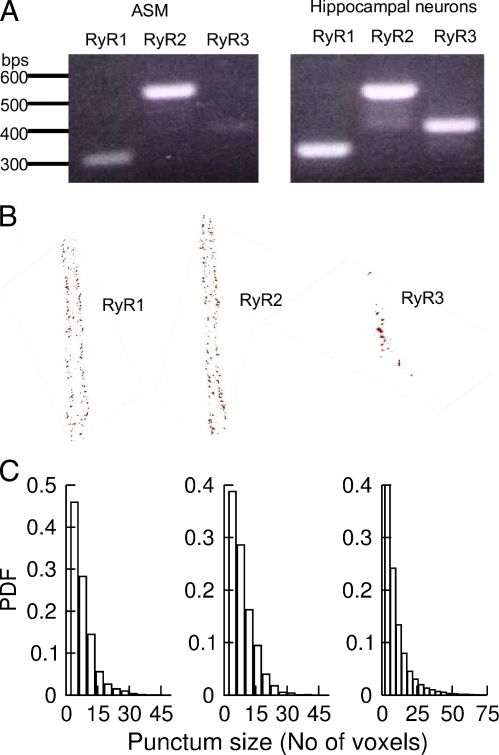
Figure 4.
Type and distribution of RYRs in mouse ASM. (A) Reverse transcription PCR detected mRNA for three types of RYRs in ASM (left) and hippocampus (right). (B) RYR1 and RYR2 localize near the plasma membrane, whereas RYR3 localizes near the nucleus. The images show the localization of three types of RYRs in a 3-D projection of 1-µm thickness in the middle of the cells. The distribution of RYRs through the whole cells can be viewed in Videos 1–4. Pixel size in x and y is 80 nm, and the z spacing is 250 nm. (C) Histograms of RYR puncta. The mean size and SEM of puncta were 6.5 ± 0.5 voxels for RYR1 (n = 5 cells), 8.2 ± 0.7 voxels for RYR2 (n = 7 cells), and 13.0 ± 1.4 voxels (n = 5) for RYR3. P < 0.09, RYR1 versus RYR2; P < 0.002, RYR1 versus RYR3; P < 0.006, RYR2 versus RYR3; unpaired t test after ANOVA. PDF, probability density function.

