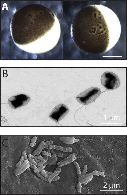
Figure 1.
Xenopus laevis oocytes infected with multi-drug–resistant bacteria. (A) Bright field micrograph of extracted oocytes showing the characteristic black foci and unpigmented halo. Bar, 0.5 mm. (B) Transmission electron micrograph of P. fluorescens cultured from compromised oocytes. (C) Scanning electron micrograph of bacteria on the surface of compromised oocytes.

