Fig. 5.

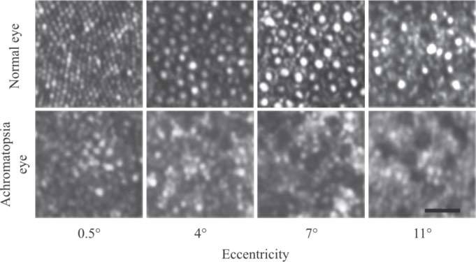
AOSLO retinal images at different eccentricities for a normal subject and a 39 year old patient with achromatopsia. The patient’s prescription on the eye imaged was −4.50sph, +0.50cyl, axis105 deg. Scale bar is 20 μm.

AOSLO retinal images at different eccentricities for a normal subject and a 39 year old patient with achromatopsia. The patient’s prescription on the eye imaged was −4.50sph, +0.50cyl, axis105 deg. Scale bar is 20 μm.