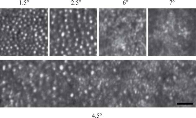
Fig. 9.

AOSLO images from an eye of a 46 year old female patient affected by AZOOR. The top images correspond to 4 different eccentricities. Images at 1.5° and 2.5° eccentricity are outside the scotoma, while images at 6° and 7° eccentricity are within it. The bottom image, centered at 4.5° eccentricity, corresponds to the edge of the area where cones are not visible. The patient’s prescription was −1.50 sph, +1.25 cyl, axis 90 deg. Scale bar is 20μm.
