
Fig. 7.
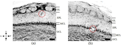
Anatomical images obtained after electrocoagulation with the site just below GL shown in (a) and the site in MCL shown in (b). The coagulation sites in the GL and MCL layers were shown as the red encircled regions. The arrows indicate respectively the anterior-posterior and dorsal-ventral parts of the rat. (a) and (b) respectively correspond to Figs. 6(a) and 6(b), and Figs. 6(c) and 6(d). Scale bars, 100 μm.