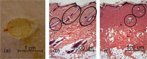
Fig. 1.

(a) Image of a 100○C, 30 second burn, along with a sample histological cross section of (b) normal and (c) burned skin confirms the existence of a 3rd degree burn. Note that post-burn edema is evident in (a) as well as the reduction in the number and size of discrete normal skin structures (marked) in burned skin relative to normal skin.
