Fig. 3.

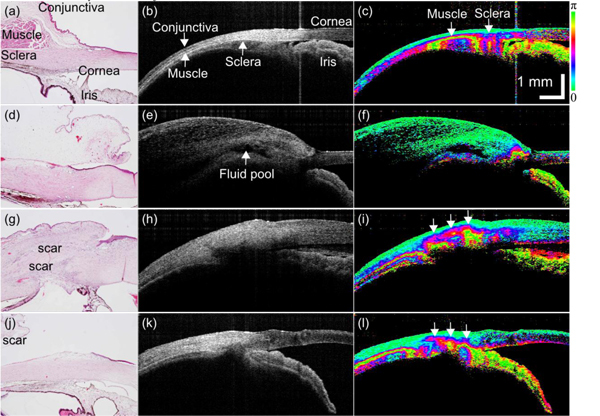
(a), (d), (g), (j) Histology images, (b), (e), (h), (k) intensity images, and (c), (f), (i), (l) phase retardation images of rabbit trabeculectomy model. (a)–(c) Control eye. Postoperative day (d)–(f) 0, (g)–(i) 8, and (j)–(l) 14.

(a), (d), (g), (j) Histology images, (b), (e), (h), (k) intensity images, and (c), (f), (i), (l) phase retardation images of rabbit trabeculectomy model. (a)–(c) Control eye. Postoperative day (d)–(f) 0, (g)–(i) 8, and (j)–(l) 14.