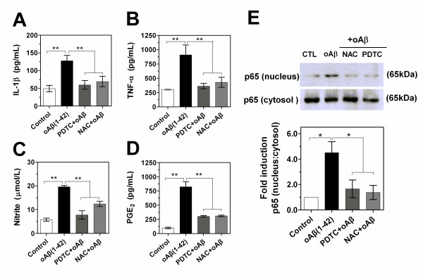
Figure 9.
Role of NF-κB signaling in the expression of inflammatory mediators in oAβ-stimulated microglial cells. (A-D) BV-2 cells were incubated with oligomeric Aβ(1-42) (1.0 μM) in the presence or absence of NAC (5.0 mM) or PDTC (20 μM). After 24 h (TNF-α, NO and PGE2) or 3 h (IL-1β), the supernatant was collected for ELISA, EIA or Griess analysis. Values correspond to mean ± S.E. (n = 4). **P < 0.01. (E) Images and quantification data of Western blot showing NF-κBp65 protein. Whole cells and nuclear extracts were prepared after pretreatment of BV-2 cells with NAC (5.0 mM) or PDTC (20 μM) for 1 h before being stimulated with oAβ(1-42) (1.0 μM) for 1 h. Cytosol NF-κBp65 was used as a protein loading for nucleus p65. Ratio of nucleus p65 to cytocol represented the level of activation of NF-κB signaling. Representative of images revealed the effects of NAC and PDTC on the activition of NF-κB. Data were expressed as relative folds compared to control. Values correspond to mean ± S.E (n = 3). *P < 0.05, **P < 0.01.

