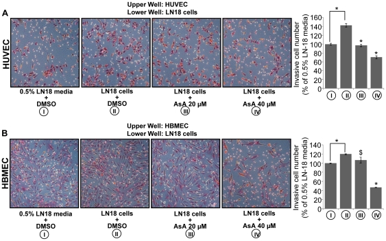
Figure 6. AsA inhibits glioma cells induced chemotactic motility of endothelial cells.
A & B. HUVEC or HBMEC (3×10⁴ per well) were seeded in the upper chamber of a Transwell plate with 0.5% serum media. In the lower chamber, LN18 cells (3×10⁴ cells per well) were treated with DMSO or AsA 20 µM in 0.5% serum media as described in ‘Materials and Methods’. HUVEC and HBMEC migrated through the matrigel layer were stained and quantified after 10 h and 22 h of their plating in the upper chamber respectively. Cell invasion data shown are mean ± standard deviation of three samples for each treatment. *, p≤0.001; $, p≤0.05.

