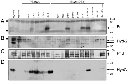
Figure 3. Western blot analysis of anaerobic enzymes in BL21(DE3).
25 µg Polypeptides in crude extracts derived from MC4100, PB1000 (Δfnr), BL21(DE3) with and without supplementation of 500 µM nickel(II)-chloride (Ni), 15 mM formate (F), 1 mM sodium-molybdate (MoO) or addition of plasmid encoded fnr (p1fnr, p10fnr, p13fnr, pCH21) and modE (p7modE) after anaerobic growth in TGYEP, pH 6.5 were separated by 10% (w/v) SDS-PAGE and transferred to nitrocellulose membranes. The samples were treated with antiserum raised against A: FNR, B: Hyd-2 (the upper arrows represents precursor and the lower arrow mature form of the Hyd-2 large subunit), C: PflB (the arrows mark the two different migrating forms typical for active protein after contact with oxygen), D: HycG (the Hyd-3 small subunit). The lane indicating the negative control contains PB1000 (Δfnr), DHP-F2 (ΔhypF), RM220 (ΔpflAB) and CP971 (ΔhycAI), from top to bottom, respectively. The asterisks signify unidentified cross-reacting species. On the right hand are given the sizes of the respective molecular mass marker (Prestained PageRuler, Fermentas).

