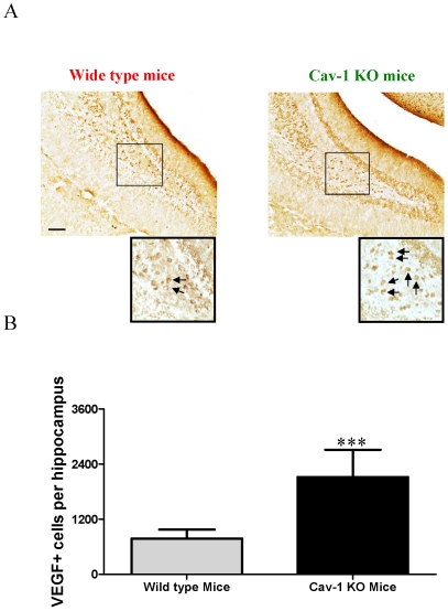
Figure 2. VEGF expression in the granule cell layer of the hippocampal dentate gyrus of wild type and Cav-1 KO mice.
A, Representative micrographs of VEGF positive cells in the granule cell layer of the hippocampal dentate gyrus of wild type mice and Cav-1 KO mice. Arrows indicate the positive cells. Scale bar = 100 µm B, Histograms showing the quantification of VEGF positive cells in wild type mice and Cav-1 KO mice (Mean ± S.D., n = 6). Wild type mice versus Cav-1 KO mice, *** p<0.001.

