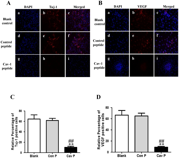
Figure 9. Effects of Cav-1 scaffolding domain peptide on the expressions of Tuj-1 and VEGF in NPCs under normoxic condition.
Cells were consistently incubated in a standard incubator with humidified 21% O2 plus 5% CO2 balanced with N2 for 14 days. Cells were cultured with fresh medium containing a synthetic cell-permeable peptide encoding Cav-1 scaffolding domain (amino acids 82 to 101, DGIWKASFTTETVTKYWFYR) or a Cav-1 scrambled control peptide (WGIDKAFFTTSTVTYKWFRY) with Antennapedia internalization sequence (RQIKIWFQNRRMKWKK) at a final concentration of 4 µM. The medium was changed every two days and lasted for 14 days. A–B. Representative immunofluorescent imaging of Tuj-1 and VEGF in NPCs at day 14. Red color: Tuj-1 and VEGF staining; Tuj-1 and VEGF staining (red color) were identified in blank control group (a, b, c), control peptide group (d, e, f) and Cav-1 peptide group (g, h, i). Nuclear localizations of Tuj-1 and VEGF were verified by co-localization with DAPI staining (blue color). C–D. Statistical analysis on the relative percentage of Tuj-1 and VEGF positive cells in NPCs (Mean ± S.D., n = 6). Blank, blank control group; Con P, Cav-1 scrambled control peptide group; Cav P, Cav-1 scaffolding domain peptide group. Cav P versus blank, ** p<0.01; Cav P versus Con P, ## p<0.01.

