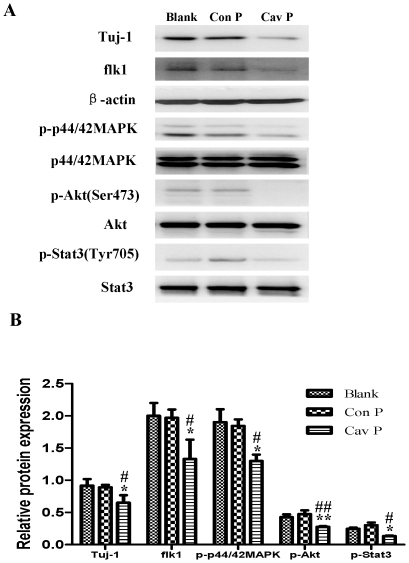
Figure 14. Effects of Cav-1 scaffolding domain peptide on the expressions of Tuj-1, flk1, p-p44/42MAPK, p-Akt and p-Stat3 proteins in NPCs under hypoxia-reoxygenation condition.
NPCs were cultured with fresh medium containing a synthetic cell-permeable Cav-1 scaffolding domain peptide (4 µM) or a Cav-1 scrambled control peptide (4 µM). For hypoxia-reoxygenation treatment, cells were exposed 1% O2 for 24 h and then switched to normoxia with fresh medium for 14 days. All data were obtained from the samples at day 14. A. Representative results of immunoblot analysis for the expressions of Tuj-1, flk1, p-p44/42MAPK, p-Akt and p-Stat3. Cell lysates were blotted with the antibodies for Tuj-1, flk1 p-p44/42 MAPK, p-Akt, and p-Stat3, in which β-actin, p44/42MAPK, Akt and Stat3 were used as internal references, respectively. Blank, blank control group; Con P, Cav-1 scrambled control peptide group; Cav P, Cav-1 scaffolding domain peptide group. B. Statistical analysis on the expressions of Tuj-1, flk1, p-p44/42MAPK, p-Akt and p-Stat3 (Mean ± S.D., n = 3). The expressions of Tuj-1 and flk1 were presented as fold activation of light units normalized to β-actin, whereas the phosphorylations of p44/42MAPK, Akt, and Stat3 were presented as the fold activations of light units normalized to p44/42MAPK, Akt and Stat3, respectively. Cav P versus Blank, * p<0.05, ** p<0.01; Cav P versus Con P, # p<0.05, ## p<0.01. Each sample was assayed at least 3 times.

