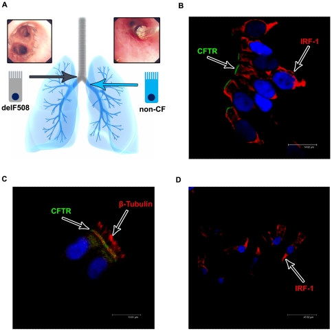
Figure 1. The post transplant lung in a CF patient; a novel way to investigate CFTR localisation.
A) Bronchial brushings (example shown in the upper right panel) from post transplant CF patients allowed both CFTR-delF508 affected epithelial cells (above the airway anastomosis from the native tissue) and non-CF epithelial cells (below the airway anastomosis from the donor lung) to be obtained from the same patient and investigate CFTR localisation and expression levels. B+C) Bronchial brushings of non-CF cells fixed in 4% paraformaldehyde and stained for CFTR (MATG1061 - FITC/Green) and IRF-1 (B) or β-tubulin (C) (TRITC/Red) with a nuclear counter stain (DAPI/Blue). CFTR is localised predominantly to the apical membrane of tall columnar epithelial cells. Images acquired on a Leica confocal microscope (×100 magnification). D) Corresponding isotype control. CFTR (MATG1061) was replaced by IgG2a at the corresponding concentrations. No non-specific staining was seen in tall columnar epithelial cells. Images acquired on a Leica confocal microscope (×63 magnification).

