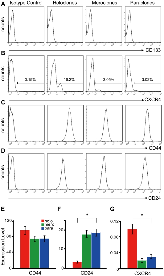
Figure 4. Expression of cancer stem cell markers in holoclones, meroclones and paraclones.
Cells isolated from holoclones, meroclones and paraclones were examined with flowcytometry for the cell surface markers of cancer stem cells. CD133 (A) was totally negative in all three types of colonies. The CXCR4+ cells (B) were detectable in all types of colonies but significantly enriched in holoclones. CD44 (C) was strongly positive and with little difference among distinct types of colonies, while CD24 (D) was expressed at a higher level in paraclones than in holoclones. mRNA level of CD44 (E), CD24 (F) and CXCR4(G) in holo-, mero-, and paraclones was also quantified with real-time PCR (GAPDH as the internal reference) and similar trends were showed (*, p<0.05).

