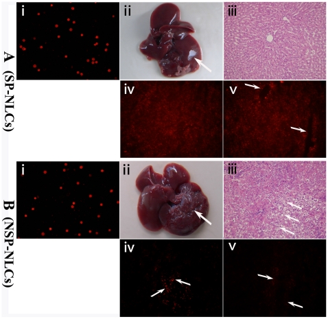
Figure 4. The regenerative effects of transplanted cells in acutely injured rats.
The membranes of (A-i) SP-NLCs and (B-i) NSP-NLCs were successfully stained with PKH26 fluorescence. After the rats were severely damaged by CCl4 and a 2/3 PH, (A-ii) transplantation of SP-NLCs enhanced liver repair (shown by a smooth surface), whereas (B-ii) the livers in the NSP-NLCs injected group still exhibit a rough surface. (A-iii) The H&E staining of livers in the SP-NLCs transplanted group. (B-iii) The livers in the NSP-NLCs injected group were stained by H&E. (A-iv) After SP-NLCs transplantation, many sporadic cells labeled by red fluorescence could be observed in the liver. (B-iv) However, minor red cells could be found in NSP-NLCs transplanted liver (arrows). (A-v) Complete hepatic cord-like structure with red fluorescence could be detected in the region near the portal area of the SP-NLCs restored liver (arrows). (B-v) Around the portal area, very weak red fluorescence in NSP-NLCs repaired liver was present (arrows). Original magnification, 200× (A, B-i, iii, iv, v). (For a better interpretation of the colored figure, the reader is referred to the web version of the article).

